
I am a political centrist. In today's polarized political climate, this means that almost all the politically opinionated people I speak with are opposed to centrism. We seem unpopular everywhere.

TROPICAL SYNAPSES
Reflections on topics including clinical neurology, recent publications in neuroscience,
philosophy of biology, "neuro-doubt" about modern media hype of new neuro-scientific procedures and methods, local oceanography, tropical horticulture, the Hawai'i health scene, and whatever else dat's da kine...
I am a political centrist. In today's polarized political climate, this means that almost all the politically opinionated people I speak with are opposed to centrism. We seem unpopular everywhere.

So the deceased's lawyer wants the brain autopsy results. Why? So that the NFL might be held liable for the brevity of a life characterized by violence?
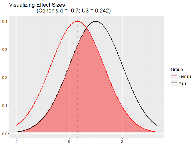
The preprint of a summary of measures of brains in 5,216 donors to the UK's Biobank program was released on bioRxiv this past week. As a summary, they found that male brains were slightly larger in most structures, on the average, than female brains. This should not be surprising given that human males are on average slightly larger than human females. After controlling for average brain volume, though, there was still frequently a 0.7 to 0.8 Cohen's D value effect size of biological gender on many brain measures, with male brains usually larger on such measures, except, as has been noted before, female brains had a slightly thicker cortex and a larger corpus callosum.
The chart above is to show the degree of overlap for such differences in an ideal normal distribution case. The charts below reflect the actual current study under review's data. It's important to note that such size differences do not generally influence social aspects of gender. For example, persons who identify themselves more with their opposite biological sex (the trans-gendered) have scans that reflect on average measures that reflect their biological sex, not their psycho-social preference for gender. A further point: variances between individuals were larger than variance between men and women, reinforcing that we need to understand a mutiplicity of each individual's own personal qualities in order to better understand them and their abilities, much more than we need to rely on a single fact such as gender. As the paper says, "Overall, for every brain measure that showed even large sex differences, there was always overlap between males and females (p. 8)."

============================================
ABSTRACT
Sex differences in the adult human brain: Evidence from 5,216 UK Biobank participants
Stuart J. Ritchie1,2*, Simon R. Cox1,2, Xueyi Shen3, Michael V. Lombardo4,5, Lianne M. Reus6, Clara Alloza3, Matthew A. Harris2,3, Helen L. Alderson7, Stuart Hunter8, Emma Neilson3, David C. M. Liewald1,2, Bonnie Auyeung1, Heather C. Whalley3, Stephen M. Lawrie3, Catharine R. Gale2,9, Mark E. Bastin2,10,11, Andrew M. McIntosh2,3, Ian J. Deary1,2
bioRxiv preprint first posted online Apr. 4, 2017; doi: http://dx.doi.org/10.1101/123729.
SEX DIFFERENCES IN THE HUMAN BRAIN
Summary
Sex differences in human brain structure and function are of substantial scientific interest because of sex-differential susceptibility to psychiatric disorders [1,2,3] and because of the potential to explain sex differences in psychological traits [4]. Males are known to have larger brain volumes, though the patterns of differences across brain subregions have typically only been examined in small, inconsistent studies [5]. In addition, despite common findings of greater male variability in traits like intelligence [6], personality [7], and physical performance [8], variance differences in the brain have received little attention. Here we report the largest single-sample study of structural and functional sex differences in the human brain to date (2,750 female and 2,466 male participants aged 44-77 years). Males had higher cortical and sub-cortical volumes, cortical surface areas, and white matter diffusion directionality; females had thicker cortices and higher white matter tract complexity. Considerable overlap between the distributions for males and females was common, and subregional differences were smaller after accounting for global differences. There was generally greater male variance across structural measures. The modestly higher male score on two cognitive tests was partly mediated via structural differences. Functional connectome organization showed stronger connectivity for males in unimodal sensorimotor cortices, and stronger connectivity for females in the default mode network. This large-scale characterisation of neurobiological sex differences provides a foundation for attempts to understand the causes of sex differences in brain structure and function, and their associated psychological and psychiatric consequences.

In the study abstract below, Kitamura and others show that long-term memory traces in the outer cerebral cortex of the mouse are laid down early in the experience to be remembered. Prior to this study the prevailing opinion was that neuronal traces for short-term memories are initially created only in the hippocampus and transferred later to long term cortex. The study below shows that initial weak traces of long term memory are created simultaneously with the strong short term memory traces in the hippocampal short term memory region, and that, over a couple weeks, the cortical area traces become stronger and more active and the corresponding hippocampal areas fade in their activity.
This suggests that prior theories were too simplistic, perhaps because they tended to model memory after human information storage, such as writing or memory disk, where information is generally laid down once and remains the same strength thereafter unless erased. Modeling the brain after our technology metaphors can be misleading.
==============================================================
ABSTRACT
Engrams and circuits crucial for systems consolidation of a memory
Takashi Kitamura1,*, Sachie K. Ogawa1,*, Dheeraj S. Roy1,*, Teruhiro Okuyama1, Mark D. Morrissey1, Lillian M. Smith1, Roger L. Redondo1,2,†, Susumu Tonegawa1,2,‡
Science 07 Apr 2017
Vol. 356, Issue 6333, pp. 73-78
DOI: 10.1126/science.aam6808
The network of memory consolidation
Memories are thought to be formed in the hippocampus and later moved to the neocortex for long-term storage. However, little is known about the mechanisms that underlie the formation and maturation of neocortical memories and their interaction with the hippocampal network. Kitamura et al. discovered that at the onset of learning, neurons for contextual fear memory are quickly produced in the prefrontal cortex. This process depends on the activity of afferents from both the hippocampus and the amygdala. Over time, the prefrontal neurons consolidate their role in memory expression. In contrast, the hippocampal neurons slowly lose this function.
Episodic memories initially require rapid synaptic plasticity within the hippocampus for their formation and are gradually consolidated in neocortical networks for permanent storage. However, the engrams and circuits that support neocortical memory consolidation have thus far been unknown. We found that neocortical prefrontal memory engram cells, which are critical for remote contextual fear memory, were rapidly generated during initial learning through inputs from both the hippocampal–entorhinal cortex network and the basolateral amygdala. After their generation, the prefrontal engram cells, with support from hippocampal memory engram cells, became functionally mature with time. Whereas hippocampal engram cells gradually became silent with time, engram cells in the basolateral amygdala, which were necessary for fear memory, were maintained. Our data provide new insights into the functional reorganization of engrams and circuits underlying systems consolidation of memory.
Normal pressure hydrocephalus (NPH) is a less common cause of dementia than Alzheimer disease, Parkinson's, or multiple stroke. Classic...


